

From Archived Tissue to Precise Molecular Answers: A Smarter Research Workflow
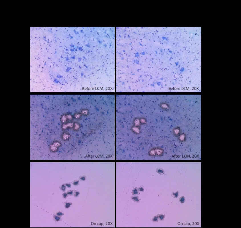
Biomedical tissue is rarely made up of one uniform cell type. Tumors, inflamed organs, and developing tissues may contain epithelial cells, immune cells, stromal tissue, blood vessels, and other structures within the same microscopic area.
This is why laser capture microdissection services can be valuable in research. They allow selected cells or tissue regions to be isolated under microscopic guidance, helping researchers focus molecular testing on a defined area rather than on a mixed bulk sample.
Start With the Right Tissue Preparation
Good results begin with proper specimen handling. Fixation, embedding, sectioning, staining, and slide preparation can all influence tissue morphology and the quality of DNA, RNA, or protein recovered later.
Researchers often work with ffpe blocks because formalin-fixed, paraffin-embedded tissue preserves architecture and supports long-term storage. These samples can be used for histology, immunostaining, in situ methods, and certain molecular studies when preparation is matched to the intended analysis.
Link Morphology With Molecular Testing
One of the strongest advantages of tissue-based research is the ability to connect visible structure with molecular information. Researchers can first identify a region by morphology, staining pattern, or marker expression and then collect that specific area for downstream testing.
This approach can be useful when comparing:
-
Tumor tissue with nearby stroma
-
Inflamed regions with unaffected tissue
-
Different anatomical compartments
-
Marker-positive and marker-negative areas
-
Distinct cell populations within one specimen
Reduce Background From Mixed Samples
Bulk extraction can sometimes hide important biological differences. A molecular signal from a small cell population may be weakened when its DNA, RNA, or protein is mixed with material from surrounding cells.
Selective collection improves sample specificity before analysis begins. Instead of relying only on an average signal from an entire section, researchers can examine a more clearly defined population and relate molecular findings to the original tissue environment.
Protect Sample Quality During Collection
Microdissection requires careful handling because collected material may be limited. Sections should provide enough detail for accurate region selection while avoiding unnecessary processing that could reduce nucleic acid or protein quality.
For RNA-focused studies, nuclease control is especially important. Researchers should also consider section thickness, staining time, storage conditions, and collection size so the captured material is suitable for the sensitivity and input requirements of the planned assay.
Match Collection to the Research Question
Not every study needs the same level of spatial precision. Some projects require a broad tissue region, while others depend on isolating a narrow population of cells. The collection strategy should reflect the biological question rather than simply collecting the smallest possible area.
A clearly defined region can improve interpretability, but overly restrictive collection may reduce yield. Balancing purity with sufficient material is therefore important, especially when downstream assays have minimum input requirements or when only a limited amount of archived tissue is available.
Plan the Downstream Assay Early
The downstream method should be selected before tissue collection begins. PCR, qPCR, sequencing, gene-expression analysis, and protein-based methods can have different requirements for sample quantity, extraction chemistry, and preservation.
Useful planning points include:
-
Expected amount of collected tissue
-
DNA, RNA, or protein as the target
-
Extraction method
-
Required controls
-
Replicate strategy
-
Storage and transfer conditions
Use Controls to Strengthen Interpretation
Well-designed controls help researchers separate biological findings from technical variation. Depending on the study, this may include matched tissue areas, positive and negative controls, replicate sections, or histologic confirmation of the selected region.
Documentation is equally important. Recording slide identity, region selection, staining conditions, collection details, and downstream processing creates a clear sample history and makes complex experiments easier to review, repeat, and compare across multiple specimens.
Build a More Efficient Tissue Workflow
A strong workflow treats histology, microdissection, extraction, and molecular testing as connected steps rather than separate laboratory tasks. Decisions made during tissue preparation can directly affect what is possible later in the study.
Researchers can improve efficiency by coordinating these stages in advance. This reduces avoidable sample loss, limits unnecessary repeat work, and helps ensure that valuable tissue is used in a way that supports the main biological question.
Conclusion
Precision tissue research depends on more than sophisticated analytical technology. The reliability of the final data also depends on how well researchers prepare the specimen, identify the relevant region, protect sample quality, and match collection methods with downstream assays.
By combining microscopic context with targeted molecular analysis, researchers can study specific cell populations with greater focus. Careful planning from tissue preparation through final testing helps make limited samples more informative and supports clearer interpretation of complex biological systems.